To observe live cells through a microscope, a sample is usually pressed onto a plate of glass. Then it remains still, and it is possible to observe the cells. The disadvantage is that this limits the behavior of the cells, and only produces 2D images.
Now researchers from the Arctic University of UiT Norway and the University Hospital of Northern Norway (UNN) have developed what they call a next-generation microscope. New technology can capture images of much larger specimens than before, while they live and work in a more natural environment.
Great development
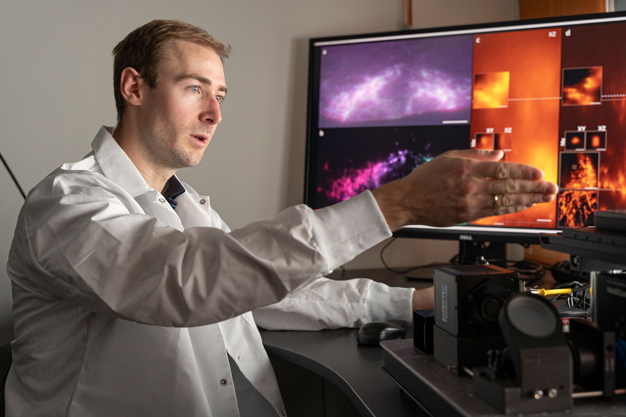
This technology provides 3D images where researchers can study the smallest details from many angles, crystal clear and visible, fully layered into different layers and where all layers are in focus.
3D microscopes do exist, but they work slowly and give worse results. The most common type works by recording pixel by pixel in a row, which are then stitched together into a 3D image. This takes time, they often can’t take more than one to five pictures a minute, and if what you’re going to take a picture is moving, it’s not very practical.
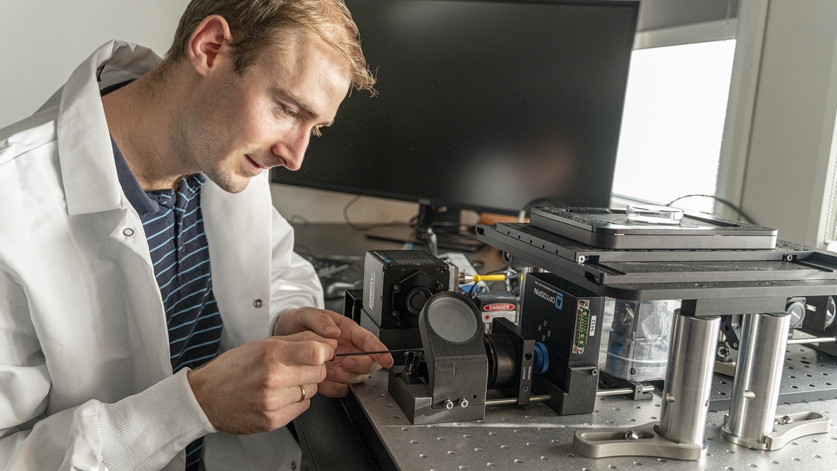
– Using our technology, we run about 100 full images per second. And we think it is possible to increase this number further, and that’s exactly what we’ve shown with our prototype, says Florian Struhl, a researcher at UiT.
The new microscope is what is called a multifocal microscope, which provides crystal clear images, completely labeled in different layers, where you can study cells from all angles.
– That’s a big deal. The fact that we can get all of this into one recording is a huge development, Strohl says.
You can see behind things
There is no question, Strohl explains, about 3D as most of us know it.
While in traditional 3D you will be able to perceive some form of depth, with the new technology you will also be able to see behind objects.
Ströhl uses an example where you see a 3D forest scene in a cinema.
– In a normal 3D image, you will see that the forest has depth, and that some leaves and trees are closer than others. With the same technology used in our new 3D microscope, you will also be able to see the tiger hiding behind the bushes. You’ll be able to see and study several layers independently of each other, Strohl says.
Now don’t use a microscope to look for tigers in the jungle, but for scientists looking for answers in the smallest detail, this can be an important tool.
The study of heart cells – as they beat
Ströhl collaborated with researchers and clinicians from the University Hospital of Northern Norway (UNN) in developing this technology.
There, they work to, among other things, understand and develop better treatment methods for various heart conditions.
Studying the living human heart is challenging, not least for technical reasons, but not least for ethical ones. Therefore, the researchers used stem cells that are manipulated so that they behave like heart cells.
In this way, they can grow organic tissue that behaves as it does in the human heart, and they can study and test that tissue to understand more of what is going on.
This tissue is roughly like a small lump of live flesh, about 1 cm in size. This creates a very demanding test situation, the heart cells are constantly beating and moving, and the sample is too large to be studied with conventional microscopes.
The new microscope handles this quite well.
– You have a piece of pumped meat in a bowl, which you want to take microscopic images of. You want to look at the smallest parts of this, and you want extreme precision. We achieved this with the new microscope, Strohl says.
Formula 1 division
Kenneth Puetz-Larsen heads a large laboratory with advanced microscopes that are used by all research groups in the UiT College of Health.
He has tested this new microscope and is optimistic.
“The concept is great, the microscope they built does things commercial systems don’t,” Larsen explains.
The lab he heads mainly uses commercial microscopes from suppliers such as Zeiss, Nikon, etc.
In addition, we have collaborations with research groups such as those represented by Florian Strohl. They’re building microscopes and testing visual concepts, they’re in a way like Formula 1 division in microscopy, Larsen says.
Larsen has great faith in Strohl’s new microscope.
Commercial microscopes should be able to be used to study all possible types of samples, while the microscope developed by Ströhl is more designed for a special task.
– Highly sensitive to light, it can shoot lotion at different points. It can work its way through the sample, and you can see a high and a low. And it happens so fast that it can be seen in practically real time. It’s a very fast microscope, Larsen says.
Tests so far show this works well, he says, and he believes this type of microscope could eventually be used on all kinds of specimens in which you look at organisms that are moving.
He also sees another advantage in the speed of this microscope.
Bright light is not “kind” to cells. Because this microscope is very fast, he explains, it exposes cells to much shorter illumination and is therefore much more careful.
He patented the technology
The prototype microscope is up and running. The researchers are now working on creating an upgraded version that is easier to use, so that more people can operate and use the microscope.
The researchers have also applied for a patent, and at the same time are looking for industrial partners who will develop this into a microscope that will be available for sale.
In the meantime, the prototype will be made available to local partners who can benefit from the new technology.
– We will also offer it to others in Norway, if they have tests that have special requirements and want to be screened, says Strohl.
You can read the research article here.
The article was first published on UiT

“Web specialist. Lifelong zombie maven. Coffee ninja. Hipster-friendly analyst.”